


Heel Spur Treatment in Singapore
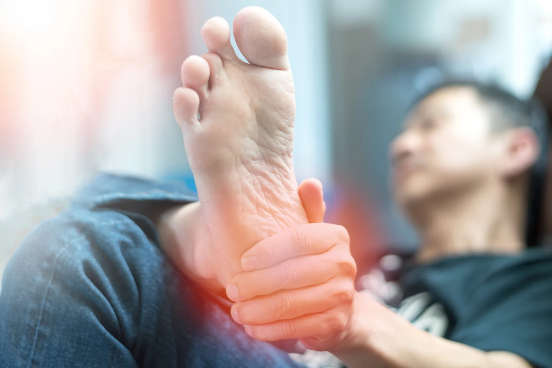
That first step out of bed in the morning should not feel like walking on broken glass. But for many people with heel spurs, it does: a sharp, stabbing pain at the base of the heel that may ease as you move, then return after long periods of sitting or standing.
Heel spur treatment at Centurion Orthopaedic Centre covers everything from conservative management to minimally invasive surgery, guided by orthopaedic surgeons in Singapore with subspecialty training in foot and ankle conditions.
What Are Heel Spurs?
Calcaneal enthesophytes, commonly known as heel spurs or bone spurs, are bony growths that form on the underside or upper backside of the heel bone (calcaneus). They develop when calcium deposits accumulate gradually over time, typically as the body’s response to repeated stress or damage to the surrounding tissues.
Heel spurs may occur on the:
- Underside of the heel at the medial calcaneal tuberosity, where the plantar fascia attaches
- At the back of the heel, where the Achilles tendon meets the bone
Foot and Ankle Conditions That May Co-Exist with Heel Spurs
Plantar Fasciitis: Chronic inflammation of the plantar fascia, a thick band of tissue that runs along the underside of the foot. This is commonly associated with heel spurs.
Achilles Tendinitis: Inflammation of the Achilles tendon, which connects the calf muscles to the heel bone. This causes pain and stiffness at the back of the heel, typically at the insertion point where a posterior heel spur may also form.
Tarsal Tunnel Syndrome: Compression of the tibial nerve as it passes through the tarsal tunnel in the ankle, causing heel pain, numbness, and tingling in the foot.
Stress Fractures: Small cracks in the heel bone caused by repetitive stress or overuse, resulting in localised pain and tenderness.
Arthritis: Inflammatory conditions such as rheumatoid arthritis can affect the joints of the foot and ankle, including the heel, causing pain and stiffness.
Heel Spur vs Plantar Fasciitis: What Is the Difference?
Bone spurs and plantar fasciitis often occur together, but they are distinct conditions that require different treatment approaches.
| Feature | Heel spur | Plantar fasciitis |
|---|---|---|
| What it is | A bony growth on the heel bone | Inflammation of the plantar fascia (soft tissue) |
| How it develops | Calcium deposited at the fascia attachment as the body attempts to repair repeated micro-trauma | Chronic stress and micro-tears in the plantar fascia at the heel bone attachment |
| Confirmed by | X-ray | Clinical examination and MRI |
| Primary cause of pain | While the spur itself is usually not the source of pain, it mechanically stretches the plantar fascia, causing pain | Yes, the inflamed fascia is typically what hurts |
| Can occur without the other | Yes, many heel spurs are asymptomatic | Yes, plantar fasciitis can exist without a visible spur |
| Treatment focus | Addressing surrounding inflammation, spur removal for persistent cases | Reducing inflammation and offloading the fascia |
If your heel pain has been diagnosed or suspected as plantar fasciitis, you can read more about our plantar fasciitis treatment in Singapore.
Symptoms of Heel Spurs
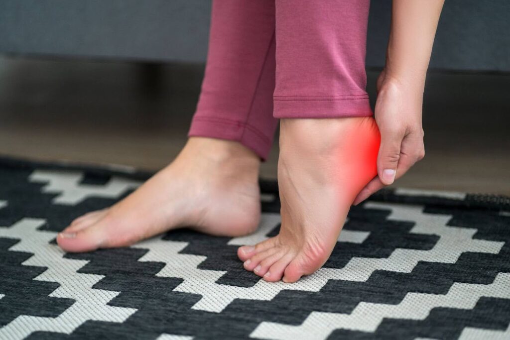
If you do have heel spurs, your symptoms may range from mild discomfort to pain that affects your ability to walk or stand. The variation is due to the location of the spur and the degree of soft tissue involvement.
Here is what you may experience:
- A sharp, stabbing pain in the heel on taking the first steps in the morning
- Pain or discomfort under the heel with prolonged standing or walking
- Pain at the back of the heel, near the Achilles tendon attachment (in cases of posterior heel spur)
- Pain that returns after long periods of sitting or rest, then eases with movement
- Chronic aching or tenderness at the bottom of the heel
- Localised swelling or inflammation around the heel
Causes and Risk Factors for Heel Spurs
Heel spurs develop gradually, typically over months or years of cumulative strain on the plantar fascia and surrounding structures.
Common causes include:
- Repeated strain on the plantar fascia: Running, jumping, or prolonged standing on hard surfaces creates micro-stress at the heel bone attachment.
- Poorly fitting or unsupportive footwear: Shoes without adequate arch support or heel cushioning increase load on the plantar fascia with every step.
- Sudden increase in physical activity: A rapid rise in training volume or intensity can overload the plantar fascia before it has time to adapt.
- Tight calf muscles or limited ankle flexibility: Reduced flexibility in the calf and Achilles complex may increase mechanical load on the plantar fascia.
- Occupation involving prolonged standing on hard surfaces: Those who work in retail, food service, or healthcare may be at higher risk.
Risk factors include:
- Flat feet or high arches: Both alter the way weight is distributed across the foot, placing higher stress on the heel.
- Higher body weight: Additional load amplifies the strain on the plantar fascia with each step.
- Age-related changes: The plantar fascia loses elasticity over time and becomes more susceptible to micro-injury.
Diagnosing Heel Spurs
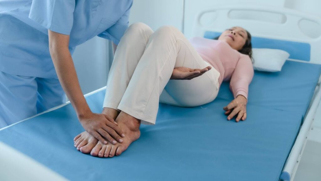
When you consult a heel spur specialist at Centurion Orthopaedic Centre in Singapore, diagnosis will typically involve a combination of clinical assessment and imaging before proceeding with bone spurs treatment.
| Diagnostic method | What it assesses | When it is used |
|---|---|---|
| Physical examination | Localised tenderness, swelling, range of motion, and weight-bearing gait | First step in all presentations; helps guide imaging decisions |
| X-ray | Confirms the presence, size, and position of a heel spur | Standard for suspected heel spur; differentiates bony from soft tissue causes |
| Ultrasound | Plantar fascia thickness, integrity, and some tendon tears | Could be used alongside X-ray, particularly when plantar fasciitis is suspected |
| MRI | Detailed view of soft tissue, muscles, and bone | Gold standard scan for plantar fasciitis and bone spur issues |
Non-Surgical Treatment Options
Mild cases of heel spurs can be managed without surgery. Non-surgical treatments focus on reducing pain and inflammation and restoring normal function. Typically, resolving the soft tissue inflammation brings lasting relief without any need to physically remove the spur. Patients often respond well to a three to six-month course of conservative treatment.
Rest and Activity Modification
| What it involves | Reducing or avoiding activities that aggravate the heel; light, low-impact movement is encouraged in the meantime |
| Who it suits | Patients in the early stages of treatment or those with a clear activity-related trigger |
| Expected outcome | Pain reduction typically within a few weeks of consistent offloading |
Footwear and Orthotics
| What it involves | Custom orthotics or insoles improve foot alignment and redistribute load away from the spur site |
| Who it suits | Patients with flat feet, high arches, or occupation-related load |
| Expected outcome | Reduced load on the plantar fascia and improved heel pain over several weeks of consistent use |
Physical Therapy
| What it involves | Structured stretching and strengthening to improve flexibility and reduce tension at the heel, ESWT and/or Ultrasound treatment |
| Who it suits | Patients at any stage of conservative management; often combined with orthotics or splinting |
| Expected outcome | Gradual improvement over four to eight weeks of consistent exercise |
Night Splints
| What it involves | Holds the foot in a gently dorsiflexed position during sleep, maintaining a mild stretch through the plantar fascia overnight |
| Who it suits | Patients with sharp, first-step morning pain |
| Expected outcome | Most patients who benefit notice a difference within three to four weeks of regular use |
Extracorporeal Shockwave Therapy (ESWT)
| What it involves | Focused sound waves delivered to the painful area, stimulating the body's natural healing response and reducing chronic inflammation |
| Who it suits | Patients with persistent symptoms that have not responded to initial conservative measures |
| Expected outcome | Gradual pain reduction over several weeks; full benefit typically apparent by eight to twelve weeks |
Corticosteroid Injection
| What it involves | Anti-inflammatory medication injected directly into the affected area |
| Who it suits | Patients with moderate to severe pain who need short-term relief to engage with physiotherapy or return to function |
| Expected outcome | Effect may be long-lasting or may be temporary |
Platelet-Rich Plasma (PRP) Injection
| What it involves | A small blood sample is centrifuged to concentrate platelets, which are injected into the affected tissue to promote tissue repair |
| Who it suits | Cases with some degree of tissue damage or tears; Patients who prefer a regenerative approach |
| Expected outcome | Benefit builds over one to three months; evidence suggests stronger long-term outcomes than corticosteroid in some studies |
Heel Spur Surgery in Singapore
Surgery is considered when pain has persisted despite adequate conservative treatment, or when the extent of structural involvement makes non-surgical management unlikely to succeed.
When is surgery recommended?
- Significant heel pain lasting more than six to 12 months despite a consistent course of conservative treatment
- Failure to respond to ESWT
- Meaningful functional limitation, such as difficulty walking, working, or engaging in daily activities, due to ongoing heel pain
- Cases where the spur is associated with a significant plantar fascia damage or structural abnormality requiring direct intervention
Types of Surgery
1. Open Heel Spur Removal Surgery
This traditional approach uses a small incision over the heel to directly access and remove the bony spur. It allows the surgeon clear visualisation of the affected area and is suitable for larger or more complex spurs.
Recovery typically involves a period of protected weight-bearing followed by a gradual return to normal activity.
2. Arthroscopic (Keyhole) Heel Spur Removal and Endoscopic Plantar Fascia Release
This minimally invasive technique uses two small incisions through which a thin camera (arthroscope) and specialised instruments are inserted. The surgeon views the heel structures in real time on a monitor, allowing precise removal of the spur and, where indicated, a partial release of the plantar fascia at its heel attachment.
The endoscopic approach means less disruption to surrounding tissue, a reduced risk of wound complications, and a faster recovery compared to open surgery.
Most procedures are performed as day surgery under regional or general anaesthesia, and patients are typically discharged the same day.
Recovery Timeline
Recovery varies by surgical approach and individual factors, but as a general guide:
| Phase | Timeframe | What to expect |
|---|---|---|
| Early recovery | Weeks 1 to 2 | Protected weight-bearing with crutches or a supportive boot; wound care and rest |
| Rehabilitation begins | Weeks 2 to 4 | Gradual increase in weight-bearing and light walking; physiotherapy typically commences |
| Walking comfortably | Weeks 4 to 6 | Most patients are walking with minimal support |
| Return to activity | Weeks 6 to 12 | Return to normal daily activities; return to sport or high-impact activity guided by your surgeon |
Meet Your Heel Spur Specialists
Whether you are exploring conservative options or considering a heel spur operation, your care is led by orthopaedic surgeons with subspecialty training in foot and ankle conditions.
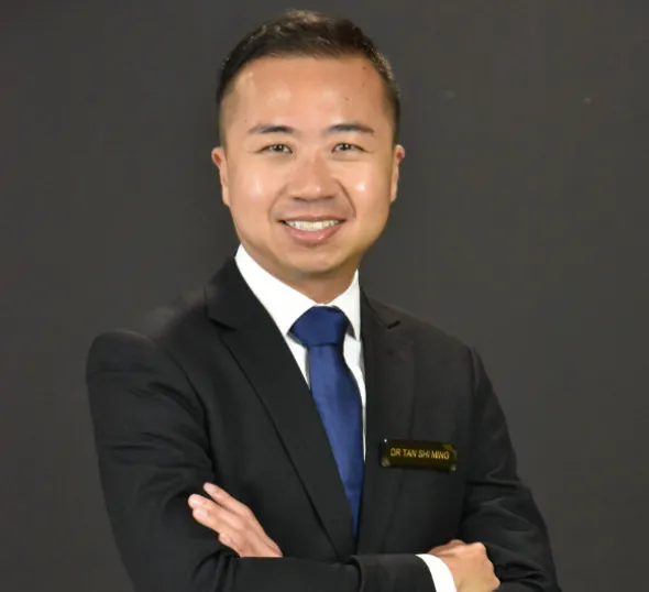
Dr Tan Shi Ming
Senior Consultant
MBBS (National University of Singapore), Fellow of the Royal College of Surgeons of Edinburgh (FRCSEd)
Dr Tan underwent subspecialty fellowship training in foot and ankle surgery in Bristol (United Kingdom), Basel (Switzerland), and Grenoble (France), under internationally recognised foot and ankle specialists. His training focused on minimally invasive percutaneous and arthroscopic techniques, complex ankle replacement, and deformity correction. He has more than 20 years of experience in orthopaedic practice.
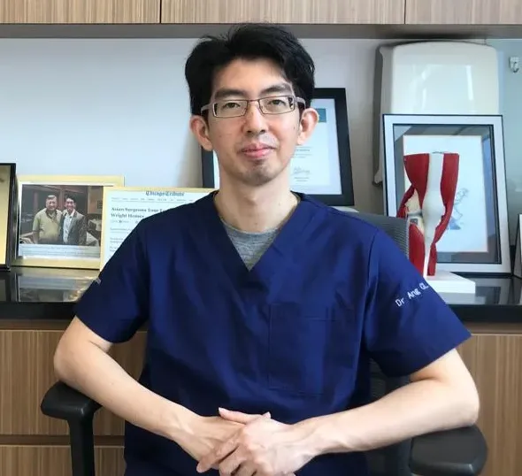
Dr Bruce Ang CL
Senior Consultant
MBBS, Fellow of the Royal College of Surgeons of Edinburgh (FRCSEd)
Dr Ang has more than 20 years of orthopaedic experience and has performed more than 6,000 orthopaedic surgeries. He has particular expertise in keyhole (arthroscopic) surgery of the knee, hip, shoulder, and ankle, as well as minimally invasive joint management and fracture care.
Prevention of Heel Spurs
Once your pain has resolved, keeping on top of a few habits can significantly reduce the risk of it returning. Here are some things you can do.
Stretch Regularly
Three stretches in particular are useful for maintaining plantar fascia and calf flexibility:
| Stretch | Starting position | How to do it | Hold and reps |
|---|---|---|---|
| Plantar fascia stretch | Seated | Cross one foot over the opposite knee, hold the toes, and gently pull them back toward the shin | 20 to 30 seconds, 3 times per foot |
| Calf stretch | Standing, facing a wall | Step one foot forward, press the back heel firmly into the ground, keep the back knee straight, and lean into the wall | 30 seconds per side |
| Towel stretch | Seated, leg extended | Loop a towel or resistance band around the ball of the foot and gently pull it toward you, keeping the knee straight | 30 seconds, 3 times |
Choose the Right Daily Footwear
In Singapore, hard-floor environments are the norm, including office buildings, shopping centres, food courts. Replace worn-out footwear regularly; cushioning degrades before the upper shows visible wear. For those who stand for extended periods at work, occupational insoles designed for hard-surface standing are worth considering.
Build Activity Levels Gradually
Avoid sharp increases in training volume or intensity. If you are returning to sport or increasing exercise, a gradual progression gives the plantar fascia time to adapt.
Cost of Heel Spur Treatment & Surgery in Singapore
The cost of heel spur treatment in Singapore varies depending on the type and duration of care required.
Non-surgical Treatment
- Physical therapy: $180 to $250 per session
- Extracorporeal shockwave therapy: $180 to $250 per session
- Corticosteroid injection: $500 to $800 per session (inclusive of consultation and procedure fees)
- PRP injection: $1,000 to $3,000 per session
Surgical Treatment
The cost of heel spur surgery depends on the surgical approach, complexity, hospital facility fees, anaesthesia, and implants or equipment used. Costs for surgical removal are variable and a consultation with our specialist is recommended to derive an estimate for your case.
MediSave and Insurance
Surgical procedures requiring hospital admission are generally claimable under MediSave. Integrated Shield Plan coverage may also apply, depending on your insurer and plan tier. Our team routinely assists patients with insurance pre-authorisation and out-of-pocket cost estimates.
For a full overview of accepted insurers, visit our Medisave and insurance page.
Why Choose Centurion Orthopaedic Centre
Subspecialty Foot and Ankle Expertise
Dr Tan Shi Ming is a fellowship-trained orthopaedic surgeon who undertook subspecialty training specifically in foot and ankle surgery across three centres in Europe. Heel spur and plantar fascia conditions fall directly within his subspecialty practice.
Full Spectrum of Care
Both conservative and surgical options are available under one roof, from orthotics and physiotherapy coordination through to keyhole surgery. Your care will be closely coordinated by our team throughout the treatment process.
Two Clinic Locations
Centurion Orthopaedic Centre operates from Mt Elizabeth Novena Hospital Specialist Centre and Farrer Park (Connexion), providing accessible appointment options across central Singapore.
Medisave and Integrated Shield Plan Acceptance
Surgical procedures are generally MediSave-claimable, and our team handles insurance coordination and pre-authorisation directly.
Bilingual Consultation
Consultations are conducted in English and Mandarin.
Frequently Asked Questions About Heel Spur Treatment in Singapore
What causes heel spurs, and how can they be treated?
Heel spurs are bony growths that form on the underside of the heel bone, typically caused by prolonged strain on the plantar fascia (the band of tissue connecting your heel to your toes), often aggravated by flat feet, tight calf muscles, or extended periods of standing. Most cases are managed with rest, physiotherapy, orthotic insoles, and anti-inflammatory medication. If symptoms persist beyond six to 12 months, procedural options such as corticosteroid injections or extracorporeal shockwave therapy (ESWT) may be considered.
Can heel spurs be treated without surgery?
Yes, the majority of heel spur cases resolve with non-surgical treatment, including physiotherapy, stretching programmes, customised orthotics, and shockwave therapy. Surgery is generally only recommended when symptoms have not improved after six to 12 months of consistent conservative management.
What is the difference between a heel spur and plantar fasciitis?
Plantar fasciitis is inflammation of the plantar fascia, while a heel spur is a bony calcium deposit that can develop as a result of that chronic inflammation. The two conditions frequently occur together, but the spur itself is often asymptomatic — plantar fasciitis is the more common source of pain. Treatment for both conditions overlaps significantly, focusing on reducing strain on the plantar fascia.
How long does recovery take after heel spur surgery?
What is the cost of heel spur treatment in Singapore?
Are heel spur treatments covered by MediSave or insurance in Singapore?
Can heel spurs come back after surgical treatment?
Recurrence is possible if the underlying biomechanical causes, such as flat feet or gait abnormalities, are not addressed. Wearing supportive footwear, continuing physiotherapy, and using custom orthotics after surgery may help reduce the likelihood of recurrence.